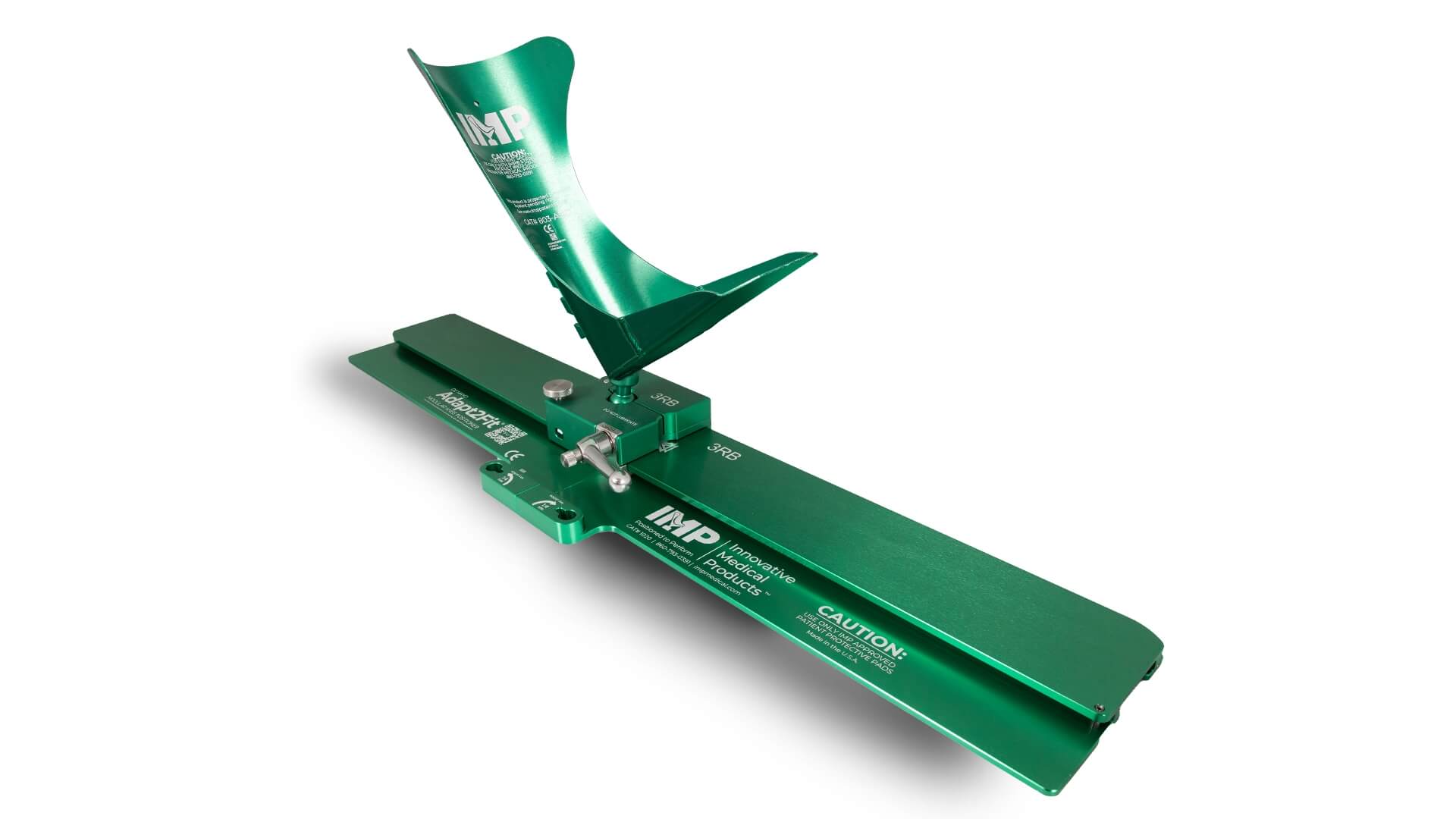
A full range of knee positioning products that reduce surgeon fatigue and assist in stabilization. Let our positioner do the heavy lifting.
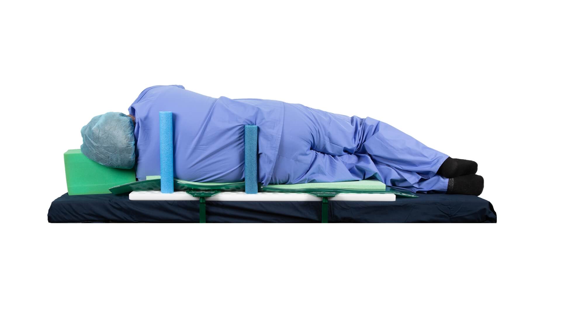
A full range of hip/lateral positioning products that accommodate a wide range of patient BMI’s. Hip surgery has never been more stable.
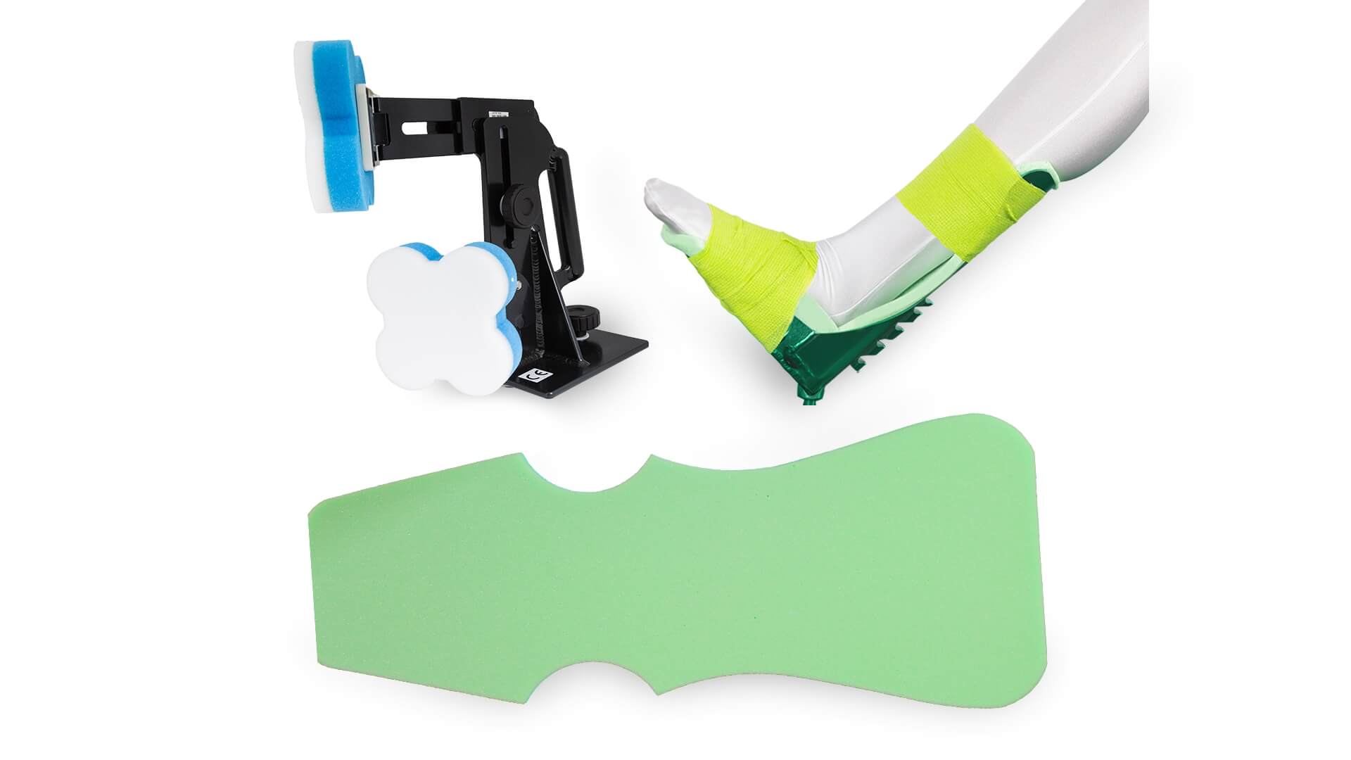
When using IMP positioners, we recommend you use the highest quality of patient pads and disposables with our products. Find your perfect foam product.
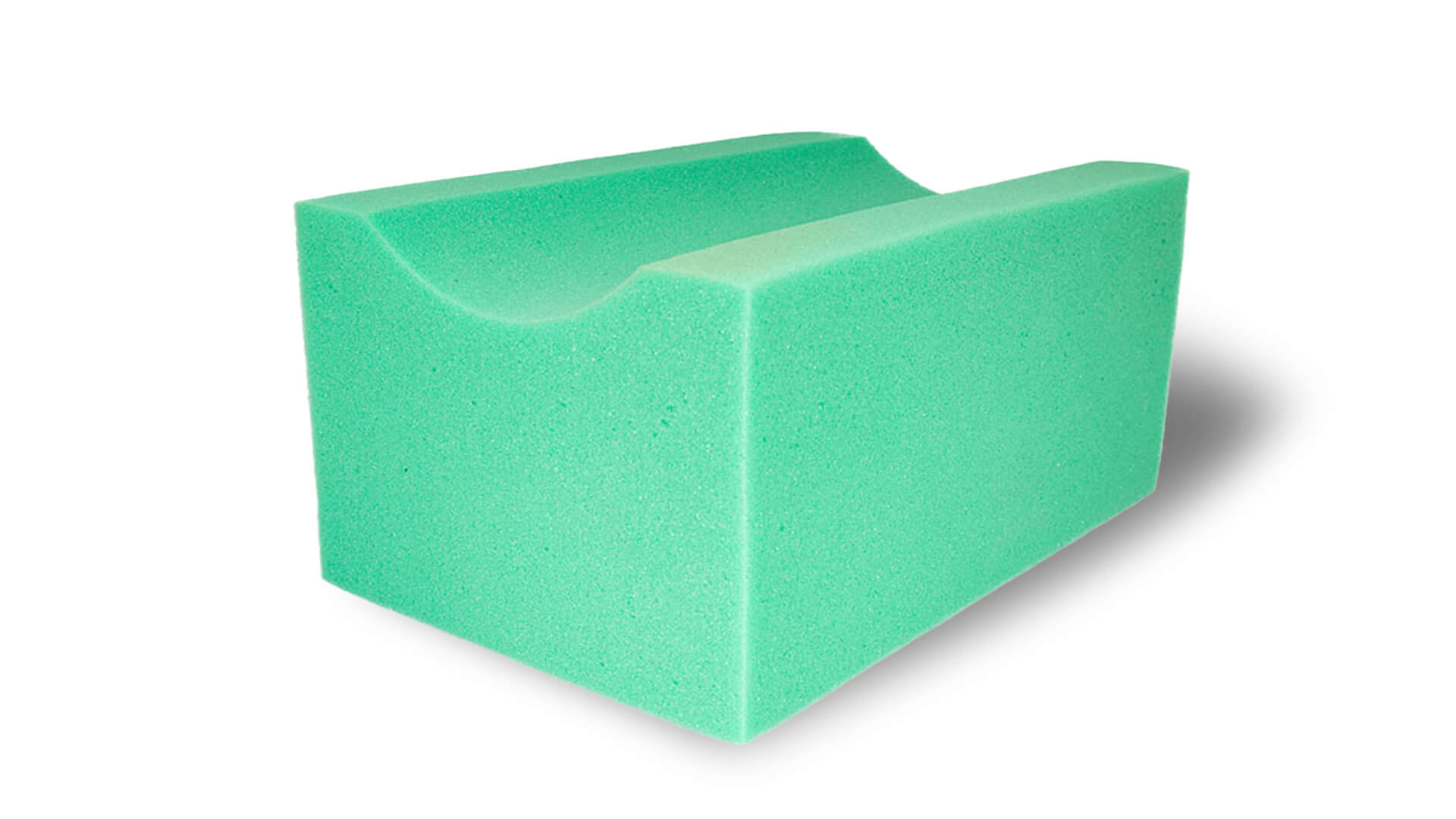
Various surgical bumps that are lint, sterile, and latex free. They have a wide range of use in the OR, and are a much safer (and cost effective) option than bundling linens.
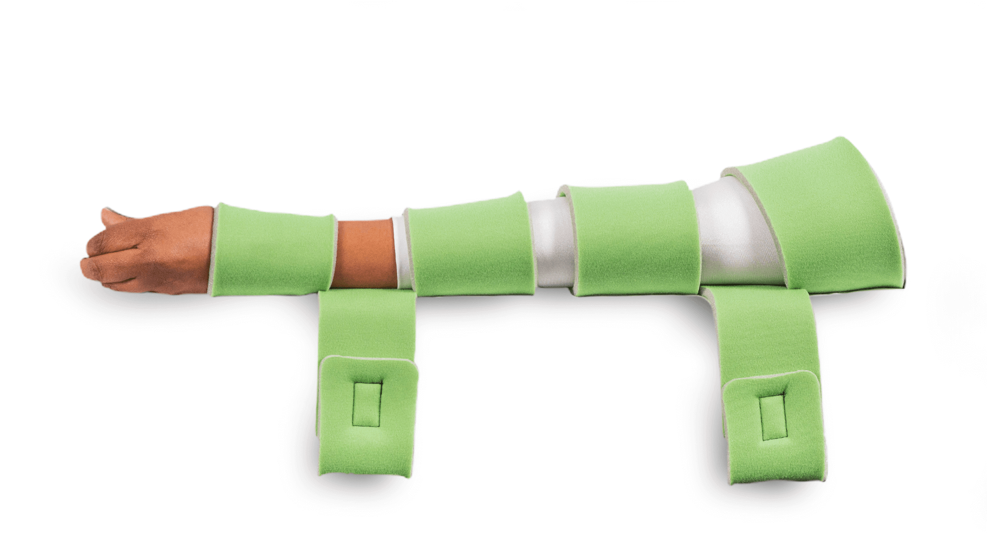
We offer more than hip and knee positioners. Check out our other various surgical positioners.
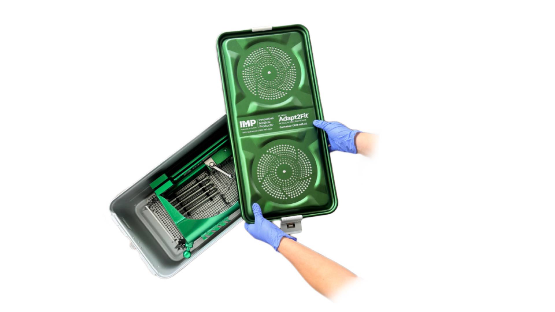
Blue wrapping large orthopedic equipment is costly, challenging, and time-consuming. Maximize efficiency and cut costs with IMP’s SteriPod™ Filtered Sterile Containers and Sterilization Case Systems.